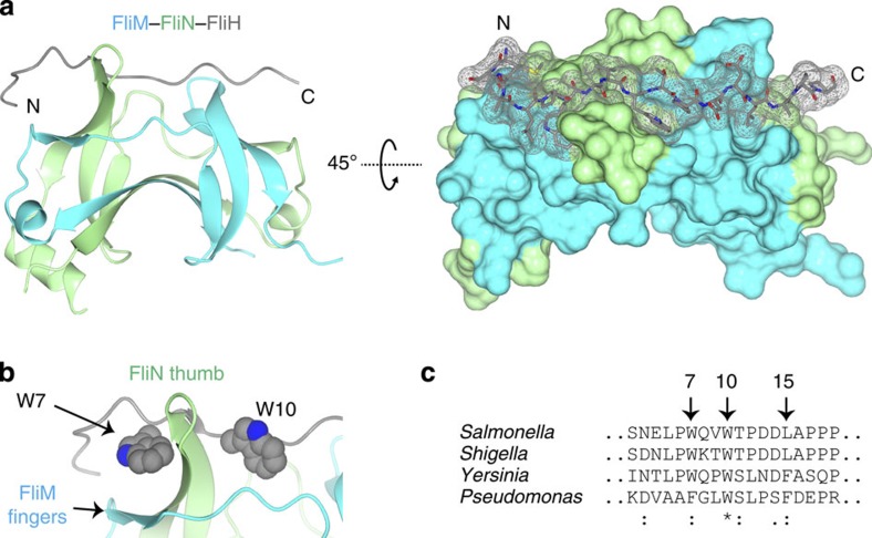
Figure 7. Structure of the SPOA1,2–APAR interaction in the flagella.
(a) Ribbon diagram (left) and surface representation (right) of the FliM–FliN–FliH structure. T4 lysozyme has been omitted. N and C indicate the amino and carboxy termini of the FliH APAR, respectively. (b) A zoomed view of the FliH aromatic clamp, with the side-chain atoms of FliH W7 and W10 represented as spheres. (c) Excerpted M-COFFEE alignment of FliH with its homologues from S. flexneri, Y. enterocolica and P. aeruginosa. Highly conserved residues of interest are noted (S. typhimurium numbering). Symbols beneath the alignment indicate the degree of conservation: asterisks denote full conservation, colons denote strong similarity, and dots denote weak similarity.

