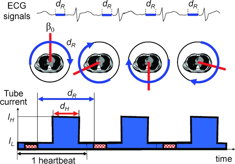
FIG. 2.
The part of projection data, dR, that corresponds to the same cardiac phase will rotate from one heart beat to another if the time durations for one gantry rotation and one heart beat are different. Images are then reconstructed from the data acquired along the blue arcs at different central angles (β0, red lines). (Bottom) Tube current modulation for cardiac CT.

