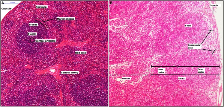
Figure 2.
Structure of spleen and lymph node. The spleen has two mayor components, white pulp that includes a central arteriole, T and B cells, and red pulp. Between red and white zone, there is a marginal zone where there are CD169+ macrophages (A). A lymph node is surrounded by a capsule, and the parenchyma is divided into cortex and medulla. The cortex has two zones: outer and inner zone. The subcapsular sinus and medulla zone contain CD169+ macrophages (B).

