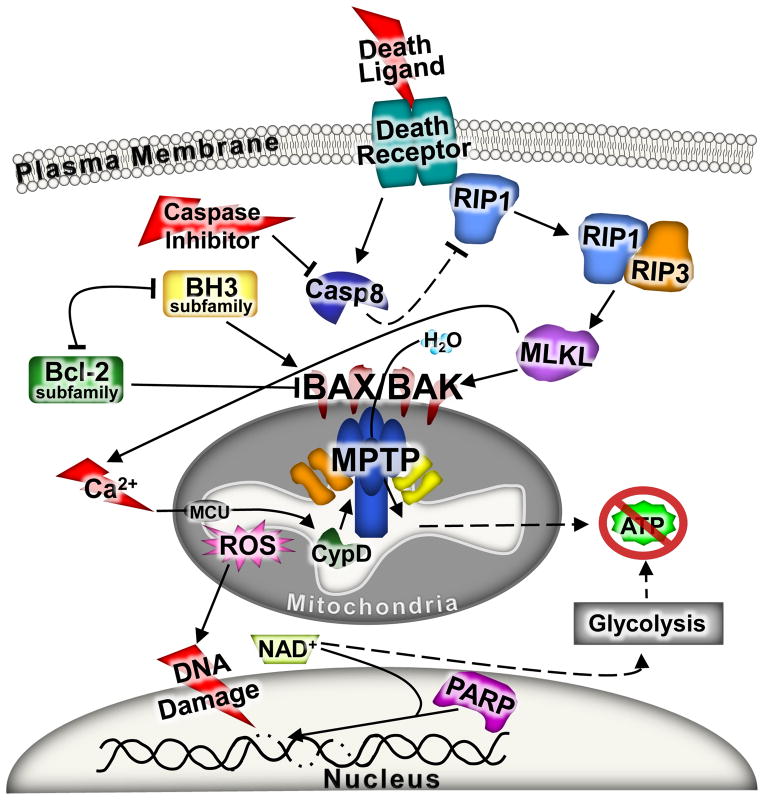
Figure 5. The unified model of regulated necrosis.
A schematic in which all three forms of regulated necrotic cell death are depicted together. Since energy depletion is a common feature for all forms of necrotic cell death, mitochondria dysfunction through MPTP opening and Bax and Bak are placed at the center of the pathway. Prior to mitochondrial dysfunction the extrinsic arm of the necrotic pathway can be engaged and it includes the RIPs and MLKL stimulated by death receptor activation with caspase inhibition. The intrinsic arm of the necrotic pathway includes stressors that increase intracellular calcium and/or induce DNA damage. Increases of calcium lead to mitochondrial dysfunction through the MPTP, while DNA damage leads to the over activation of PARP. Both of these later mechanisms result in dysfunctional mitochondria and a further exacerbation of the injury events and culmination in necrotic cell death. Due to the rapid decline of ATP generation and the rupture of mitochondria the cell loses the ability to regulate plasma membrane integrity and necrosis fully ensues.

