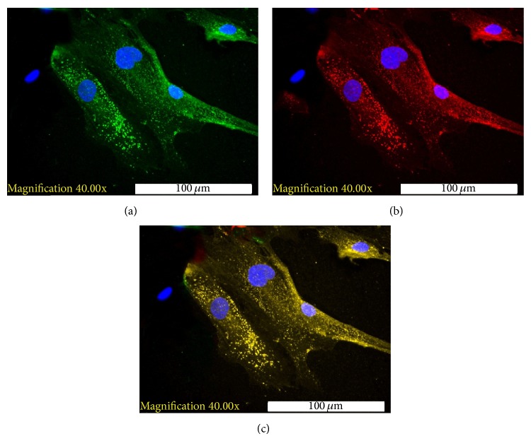
Figure 1.
Immunofluorescence staining of differentiated HBM-MSCs ((a) selected field). (a) Positive staining for intracytoplasmic insulin granules (green) with counterstaining for DAPI (blue). (b) Positive staining for c-peptide (red) with counterstaining for DAPI (blue). (c) Electronic merging of the insulin and c-peptide staining. The coexpression of insulin and c-peptide (yellow) was detected in the same cells.

