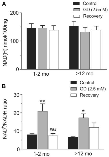
Fig. 3.
Effect of glucose deprivation (2.5 mM) on total NAD(H) content (A) and the NAD+/NADH ratio (B) in hippocampal slices of young adult (1–2 months) and aging rats (12–20, and >22 months). (A) Total NAD(H) content remained stable in hippocampal slices after 40 minutes 2.5 mM glucose condition and 40 minutes 10 mM glucose recovery. (B) The NAD+/NADH ratio increased significantly after moderate glucose deprivation (2.5 mM) but recovered completely after 40 minutes of 10 mM glucose in young slices. In contrast, the NAD+/NADH ratio remained relatively oxidized in hippocampal slices from aging rats after return to 10 mM glucose compared with the control levels (*p < 0.05, **p < 0.01; GD vs. control. ###p <0.01; recovery vs. GD; 2-way ANOVA followed by Sidak multiple comparisons test, n = 6 [young] and 5 [aging]).

