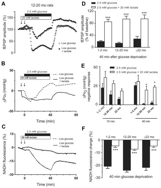
Fig. 5.
Lactate supplementation improves neuronal dysfunction induced by moderate glucose deprivation in hippocampal slices across the life span. (Left) Representative traces of fEPSP (A), NADH response (B), and tissue Po2 (C) recorded in hippocampal slices from12- to 20-month-old animal exposed to 2.5 mM glucose with or without (±) lactate 20 mM. (Right) Data summary: (D) lactate supplementation significantly prevented the suppression of fEPSP during low-glucose condition. (E) Lactate increased the rate of oxygen utilization (F) and prevented the decline of NADH fluorescence in aging rats. (*p < 0.05, ***p < 0.001; 2.5 mM glucose + lactate vs. 2.5 mM glucose; 2-way ANOVA followed by Sidak multiple comparisons test; data are mean ± SEM [n = 5]). Abbreviations: ANOVA, analysis of variance; fEPSP, field excitatory postsynaptic potential; SEM, standard error of the mean.

