
Figure 2. Turducken model of mitochondrial structure and fusion by Mfn1, Mfn2, and Opa1.
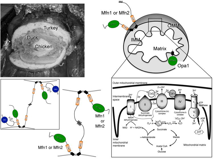
Top, multi-compartment structure of turducken (left) and cartoon mitochondrion (right). Outer mitochondrial membrane (OMM) fusion protein mitofusins (Mfn) 1 or 2 and inner mitochondrial membrane (IMM) fusion protein Opa1 are shown with GTPase domains in green. Exploded view of electron transport chain and associated pathways is bottom right. Bottom left, schematic depiction of Mfn-Mfn binding in trans, tethering two mitochondria; inset is authors’ conception of how pro-fission protein Drp1 (blue) may facilitate Mfn-mediated fusion.