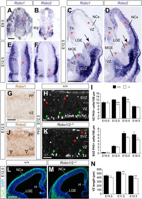
Figure 2. Robo1 and Robo2 Are Expressed in CNS Progenitors and Are Required to Sustain Ventricular Mitosis.
(A–F) Coronal sections through the spinal cord (A and B), telencephalon (C and D), and thalamus (E and F), showing expression of Robo1 and Robo2 mRNA at the indicated ages. Arrows point at the pallial-subpallial boundary. Red asterisks mark progenitor regions.
(G and J) Immunohistochemistry for Robo1 and Robo2 in the E12.5 NCx. (H and K) PH3 stains in the E12.5 neocortex of control and mutant embryos. Green arrowheads indicate VZ mitoses; red arrowheads indicate SVZ mitoses. (I) Quantification of linear density of PH3+ nuclei in the VZ and SVZ of controls (+/+) and Robo1/2 mutants (-/-) at different stages; mean ± SEM (n = 4–6 embryos per group).
(L and M) TuJ1/DAPI stains in the E12.5 NCx of control and mutant embryos.
(N) Quantification of the length of the pallial VZ, as indicated by the dotted lines in (L) and (M). Mean ± SEM (n = 4–7 embryos per group). t test; **p < 0.01; ***p < 0.001.
Scale bars equal 50 μm (A and B), 500 μm (C–F, L and M), and 100 μm (G–K). drg, dorsal root ganglion; fp, floor plate; PP, preplate; rp, roof plate; zli, zonal limitans intrathalamica. See also Figures S2 and S3.

