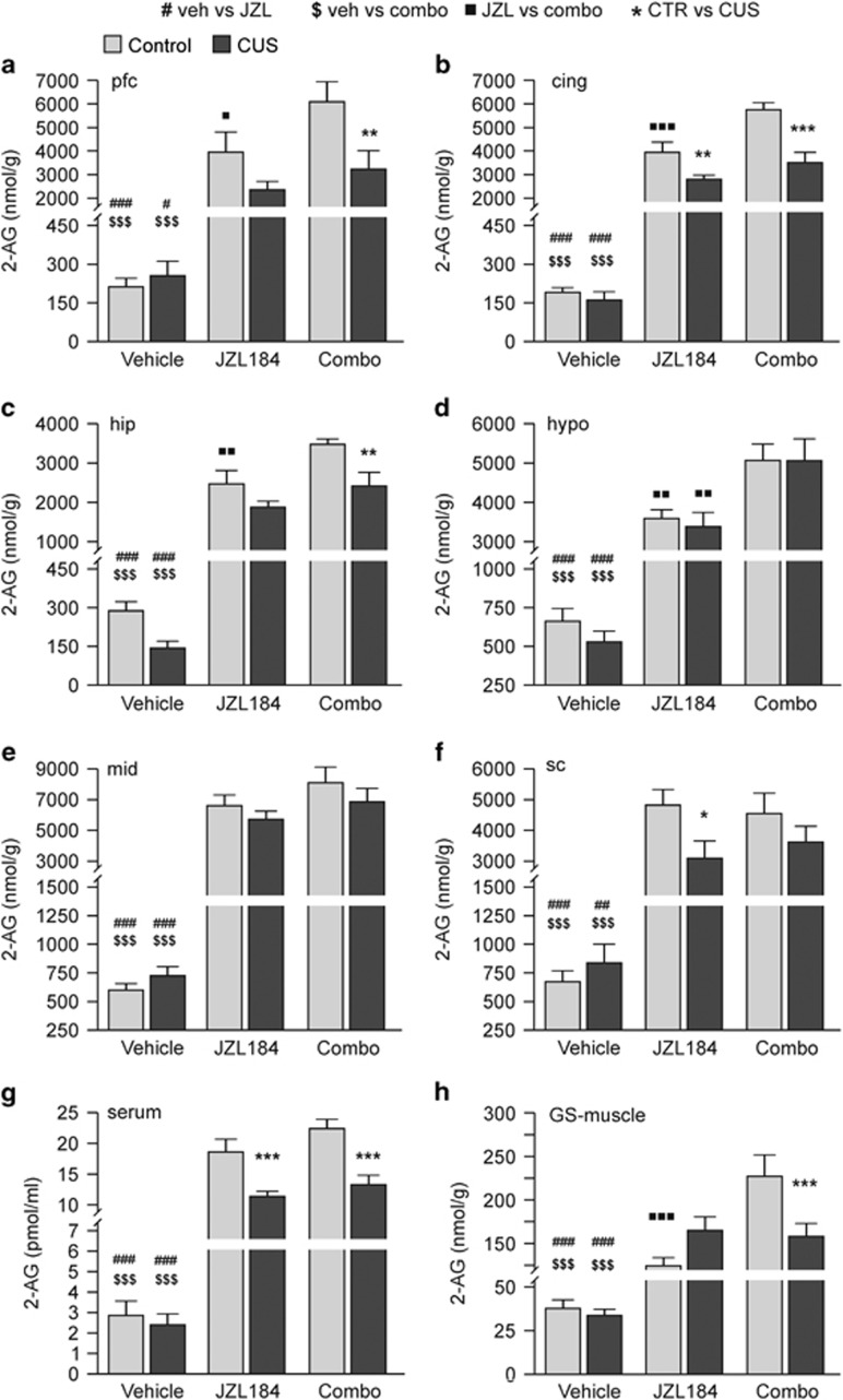
Figure 5.
Quantification of 2-arachidonoylglycerol (2-AG) in brain, muscle, and serum by LC-MRM. CUS did not change 2-AG levels in brain (a–f), serum (g), and left GS muscle (h), although a non-statistically significant decrease (p=0.2) of 2-AG in vehicle-treated CUS mice compared with controls was observed only in hippocampus (c). JZL184 treatment induced a ∼3-fold increase in 2-AG levels in all brain regions examined (a–f) of both animal groups. A statistically significant difference between JZL184-treated control and CUS mice was found in cingulate cortex (b), spinal cord (f), and serum (g), indicating that the effects of JZL184 were stronger in control than in CUS mice. Combo treatment induced a statistically significant increase in 2-AG level compared with vehicle-treated animals in all tissues examined (a–h). Compared with JZL184-treated mice, combo treatment induced a synergistic increase in 2-AG level only in control mice in almost all tissues (a–d and h) except midbrain (e), spinal cord (f), and serum (g). In CUS mice combo-induced synergism occurred only in hypothalamus (d). Statistical differences between specific groups are shown on each bar. #, $, , *p<0.05; ##, $$,
 , **p<0.01; ###, $$$,
 , ***p<0.001, Bonferroni's multiple comparison tests after significant two-way ANOVA; n=8–10 animals in each group. Additional statistical analyses are reported in Table 4. 2-AG, 2-arachidonoylglycerol; cing, cingulate cortex; hip, hippocampus; hypo, hypothalamus; GS-muscle, gastrocnemius-soleus muscle; mid, midbrain; pfc, prefrontal cortex; sc, spinal cord.

