Figure 5.

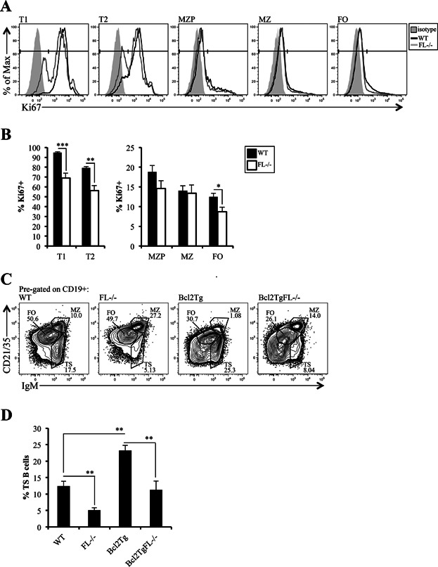
Proliferative capacity, but not survival is impaired in TS B cells in FL-/- mice. (A) Intracellular flow cytometric analysis of Ki67 in T1, T2, MZP, MZ, and FO B cell subsets. The filled histogram represents the wild-type (WT) isotype control; the black line indicates Ki67 staining in T1, T2, MZ, MZP, or FO subsets in WT, and the gray line for FL-/- mice. FL-/- isotype and WT and FL-/- unstained controls looked identical to WT isotype control staining (data not shown). T1, T2, MZ, and FO B cell subsets are immunophenotypically defined in Figures 1 and 3. MZP are CD19+ CD21/CD35hi IgM+ CD23+. (B) Bar graphs depicting the frequency of Ki67+ cells in WT and FL-/- T1, T2, MZP, MZ, and FO B cells. The bars represent WT (black) or FL-/- (white). (A and B) Data are representative of 11 mice/genotype and four independent experiments. (C) Flow cytometric analysis (pre-gated on CD19+) to examine TS, MZ, and FO B cell subsets in spleens of WT, FL-/-, Eu-Bcl2Tg (Bcl2Tg), and Eu-Bcl2Tg FL-/- (Bcl2TgFL-/-) mice. (D) Bar graph illustrating percentages of TS B cells across the four genotypes displayed in (C). (C and D) Data are representative of 5–8 mice/genotype and four independent experiments. (B and D) Error bars represent mean ± SEM. *, **, and *** represent statistically significant differences measured using the Student's t-test at P ≤ 0.05, P ≤ 0.005, and P < 0.0001, respectively, between the means of different genotypes.
