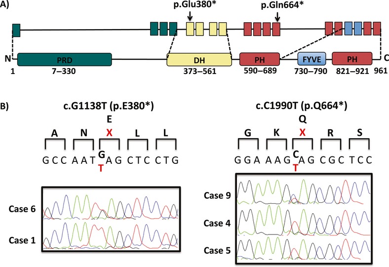
Figure 2.

Detection of FGD1 mutations. (A) Schematic representation of the domains of the FGD1 protein showing mutations (p.Glu380* and p.Gln664*) identified in patients with AAS. Arrows indicate the positions of the mutated nucleotides in FGD1. (B) sequencing results (p.Glu380* and p.Gln664*) detected in exon 5 and 12, respectively. The altered amino acids are shown in red.
