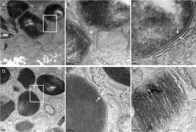
Fig 4. Identification of F. tularensis in AT-II cells.
TEM images of infected mouse lungs at 24 hours post infection. To differentiate between lamellar bodies and bacteria in AT-II cells, infected cells were observed under high magnification. Lamellar bodies were classified as dark stratified stained organelles whereas; bacteria were identified by smooth dark staining that lacked stratification. Panel A—Cell containing a F. tularensis organism magnified 8,000-fold. White rectangle marks area of interest that is shown in panels B and C. Panel B is the area of the white rectangle in Panel A magnified 25,000-fold. A bacterium and the membrane surrounding the organism are shown. Panel C—The white arrows are pointing to the double membrane (outer and inner membrane) of F. tularensis. (Panel D) An intracellular organism magnified 8,000-fold. The white rectangle marks an area of interest shown in Panel E. Panel E is the area of the white rectangle in Panel D magnified 25,000-fold. Panel F—25,000-fold magnification of a lamellar body demonstrating the stratified staining from the packaged surfactant.

