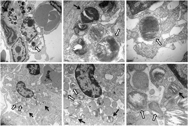
Fig 6. TEM of F. tularensis in dying AT-II cells in vivo.

TEM images of infected mouse lungs at both 32 and 48 hpi infected with Schu S4. (A-C) AT-II cells containing Francisella going through cell death characterized by lack of electron density of the cytosol and swollen mitochondria. (D-F) A separate AT-II cell undergoing cell death infected with Francisella. The open arrows identify the bacteria in each field and the solid black arrows indicate lamellar bodies in the cells.
