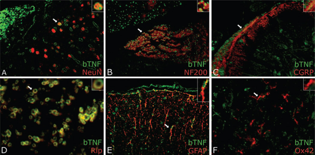
Figure 3.
Biotinylated tumor necrosis factor-alpha (TNF-α) detected using neutravidin-fluorescein isothiocyanate (FITC) (green) in the spinal cord dorsal horn ipsilateral to sciatic CCI nerve injury 96 h after intra-sciatic injection. Sections were dual-labeled with neuronal marker NeuN (A) for large neurons, NF200 (B) for small neurons, calcitonin gene-related peptide (CGRP) (C) for CGRP+ fibers, Rip (D) for oligodendrocytes, glial fibrillary acidic protein (GFAP) (E) for astrocytes, or Ox42 (F) for microglia. TNF-α colocalized (yellow) with all markers except Ox42. Magnification 1200× (A, C, D) or 600× (B, E, F).

