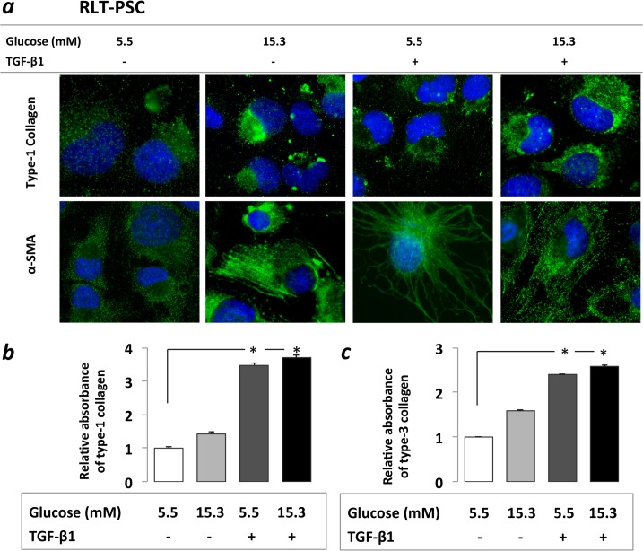
Fig 2. Immunocytochemistry (a) and ELISA assessment (b) of PSC activation.
Increased α-SMA and type-1 collagen protein expression was found on immunocytochemistry after PSCs were exposed both to 21 days of hyperglycemia or 48h of TGF-β1 (a). Increased type-1 and type-3 collagen levels were observed in PSC cell culture supernatant in the ELISA studies, however the increases were statistically only significant after the TGF-β1 treatments both with and without prior CHG exposure, but not after CHG exposure alone (b). Significant differences (p<0.05) are indicated *

