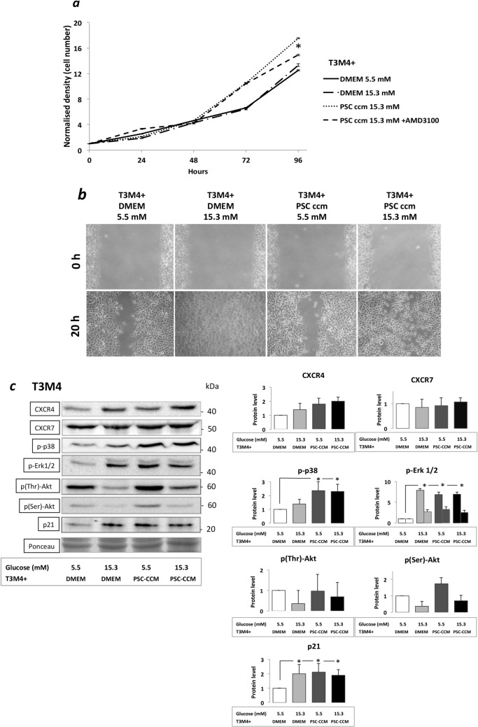
Fig 5. Proliferation and migration assays of T3M4 cells and the Western blot analyses of key signaling molecules: (a) Sulphorodamine B test.
Proliferation of T3M4 cells incubated with different conditioned PSC culture medium and the CXCR4 inhibitor AMD3100 after 24, 48, 72 and 96 hours of treatment. T3M4 cell proliferation (solid line; untreated control) was increased significantly after incubation with PSC supernatant (dotted line) provided that PSC were prior exposed to CHG. The presence of the CXCR4 inhibitor AMD3100 (dashed line) could partially antagonize this proliferation induction after 96h. (b) Migration of T3M4 cancer cells in a wound-healing assay. Substantial effect of hyperglycemia on the migration of cancer cells: a rapid direct effect of elevated glucose level was found. There was no striking effect of conditioned PSC culture mediums on T3M4 cell migration. (c) Western blot analyses of key signaling molecules in T3M4 cells. Cancer cells were exposed to hyperglycemia or different conditioned PSC culture medium. Best representative images of key signaling molecules of the cancer cells are indicated in WB membranes. Ponceau staining was used to assess the equal loading of gels. Significant (p<0.05) differences are indicated.*

