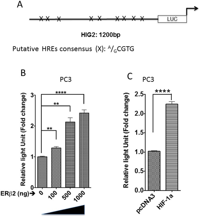
Fig 2. HIG2 promoter is activated by ERβ2 expression.
A. Schematic outline of the HIG2 promoter construct where (X) is representing sequence of HIF-1α response elements (HRE). B. Increasing amount of transient transfected ERβ2 expression plasmid into PC3 cells successively activates the transiently transfected HIG2 promoter as shown by luciferase assay of HIG2-A-LUC. The graph shows the data as an increase in luminescence of cells transfected with increased concentration of ERβ2 (mean of three separate experiments (±s.e.m.) calculated using Student’s t-test, ***p≤0.0002). Comparison of concentrations—0μg to 1000 μg. C. Addition of expression plasmid for HIF-1α activates HIG2 promoter in PC3 cell lines. The graph shows the data as an increase in luminescence of cells transfected with HIF-1α (mean of three separate experiments (±s.e.m.) calculated using Student’s t-test, ****p≤0.0001).

