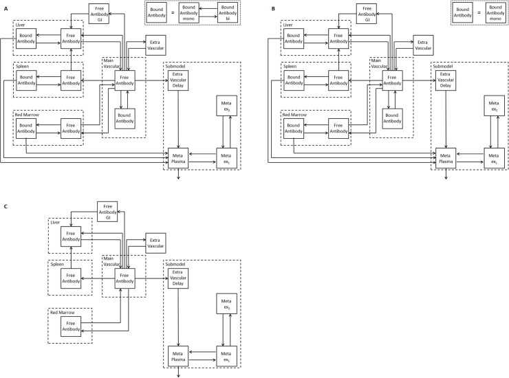
Fig 1. PBPK model for radiolabeled anti-CD66 monoclonal antibodies.
Models for (A) fully intact (both antigen-binding sites are active, i.e. bivalent binding of antibody possible), (B) half (one antigen-binding site is active, i.e. monovalent binding) and (C) non-immunoreactive antibody (both antigen-binding sites are inactive, i.e. no binding). Due to the equivalence of both valences of the antibody, the fractions of antibodies in (A), (B) and (C) are determined as follows: With the probability rim of one antibody valence being immunoreactive, the fractions of fully, half or non-immunoreactive antibody injected in (A), (B) and (C) are , and (1 − r im)2, respectively. The model consists of two equal subsystems describing the biodistribution of the labeled and unlabeled antibodies (this is true for A, B and C). The labeled and unlabeled species are competing for binding to free antigens (only A and B). The subsystems are additionally connected via physical decay, i.e. when the radiolabel decays the molecule enters the corresponding unlabeled compartment. The corresponding model equations are provided in supplement S1 Text. Radiolabeled and unlabeled antibodies are intravenously injected (main vascular compartment). The antibodies are distributed via blood flow to the main CD66 antigen expression sites. The discontinuous capillary structure of the liver, spleen and the red marrow allows the modeling of the vascular and interstitial space as one compartment. The degradation rate of bound antibody is assumed to be the same in all organs. The submodel for degraded antibody is adopted from Houston et al. [3, 28]. GI = gastrointestinal tract; Meta = metabolites in plasma; ex1, ex2 = extravascular metabolites; mono = monovalent and bi = bivalent binding.

