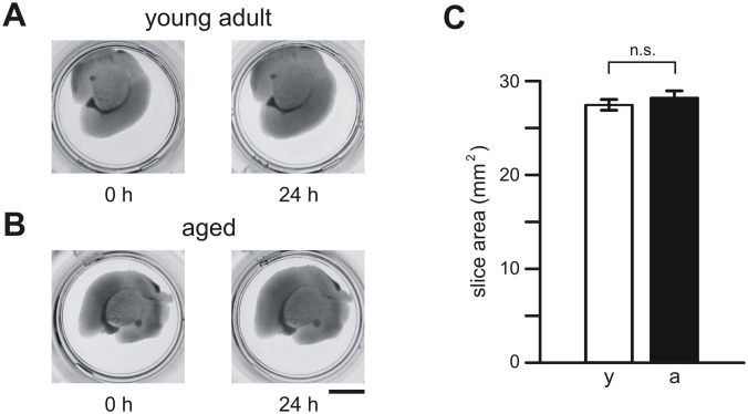
Fig 1. Brain slices from young adult and aged mice.
(A) Pictures of individual brain slices from young adult (A) and aged (B) mice. Slices placed onto culture inserts in 24-well plates are shown immediately (0 h) and 1 day (24 h) after slice preparation. (C) Mean values (±SEM) of brain slice area determined for young adult mice (y, open bars) and aged mice (a, closed bars). Calibration bar: 5 mm; n.s., not significantly different.

