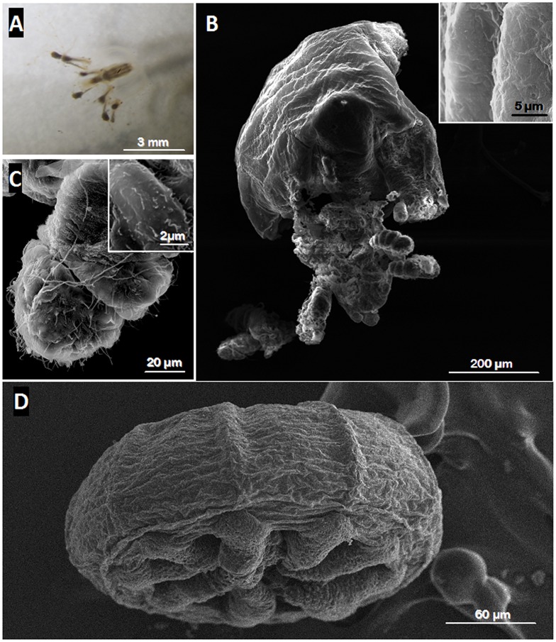
Fig 5. Ultrastructural analysis of a Medusa.
Light micrograph of the medusoid stage of Cladonema sp. (A) shows that the tentacles extended around the bell while SEM image (D) shows that the tentacles were retracted due to the chemical fixation artefact. No bacterium was observed on the bell surface of the medusa (B, D) or on its tentacles (C-D). The two insets clearly confirmed the absence of ectosymbiotic bacteria on the bell surface (B) and on the tentacles (C) of the medusa.

