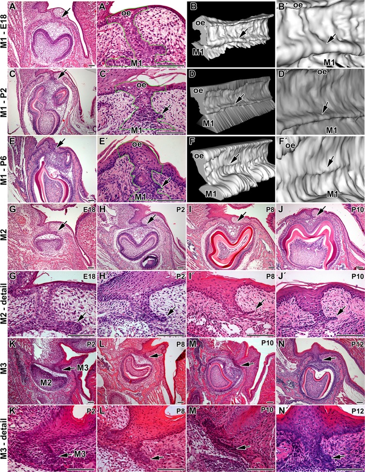
Fig 1. Dental lamina morphology in monophyodont mouse.
A: Dental lamina in monophyodont mouse at E18 is composed of the dental stalk area (ds) connecting the first lower molar (M1) to the oral epithelium and rudimental successional dental lamina (arrow). Dental stalk is very short and tooth develops in the close proximity to the oral epithelium. A´: Detail of dental stalk area at E18. B: 3D reconstruction of the dental stalk area as inclined view from rostral part. B´: Detail of continuous rudimental successional dental lamina (arrow) at E18. C: Lower power of the first molar and detail of the dental stalk area (C´) at P2 with smaller successional dental lamina. D, D´: Rudimental successional dental lamina (arrow) is smaller at P2 especially at rostral and caudal end of the first molar (M1) as shown by 3D reconstruction. E, E´: Dental stalk area at P6. F, F´: The size of successional dental lamina is much smaller at P6 and it forms distinct structure just at cusps level (arrow). G, G´: The successional lamina is protruding on the lingual aspect of the second molar (M2) at E18. H, H´: The rudimental successional dental lamina of M2 became smaller at P2 and P8 (I, I´). J, J´: In contrast to the first molar area, the lamina is still visible close M2 at P10. K, K´: There is no successional lamina close the third molar at P2 as the teeth reached just early cup stage. L, L´: In the area of the third molar (M3), successional lamina was well visible at P8. M, M´: Later in development became thinner. N, N´: At P12, it was still observable at P12 in contrast to the first and the second molar area. arrow—rudimental successional dental lamina, oe—oral epithelium. Hematoxylin-Eosin. Scale bar—100 μm

