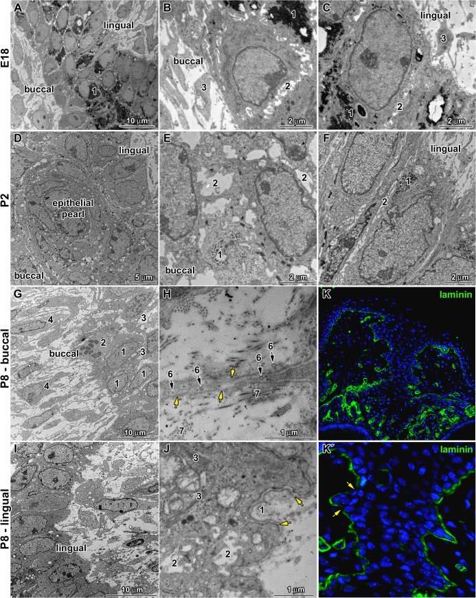
Fig 4. Differences between buccal and lingual parts of the dental stalk.
A: Low power of the dental stalk area at E18. Central area contains large amount of glycogen (1). B: Detail of buccal side of the dental stalk at E18 with glycogen (1) in cytoplasm of deeper cells, expanded intercellular spaces (2) among epithelial cells and mesenchymal cells (3) in close proximity to basement membrane. C: Detail of lingual side of the dental stalk at E18 with high cylindrical cells separated by tight intercellular spaces (2) among epithelial cells. Large amount of glycogen (1) is located also in the basal layer of buccal cells. D: Low power of the dental stalk area at P2 with epithelial pearl in the central area. E: Detail of buccal side of the dental stalk at P2 with lower amount of glycogen (1) in the epithelial cells and extended intercellular spaces among them (2). F: Detail of lingual side of the dental stalk at P2 with reduced amount of glycogen (1) in contrast to previous stage as well as very tight intercellular spaces (2). G: Low power of the buccal side in the dental stalk area at P8. Epithelial cells (1) are separated by large intercellular spaces (3). Only occasional cells in mitosis (2) are observed. Long cytoplasmatic processes of fibroblasts (4) are oriented towards the epithelium. H: Detail of buccal side at P8, where epithelial cells send extremely long cytoplasmatic processes with numerous vesicles (6) out into the connective tissue. Basement membrane (yellow arrow) is surrounded by collagen fibers (7). I: Low power of the lingual side in the dental stalk area at P8. J: Higher detail of lingual side at P8 on basement membrane (yellow arrow) surrounded by large amount of extracellular matrix (1). Intercellular spaces (2) between epithelial cells contain numerous short cytoplasmatic processes. Bundles of intermediate filaments (3) are visible in the basal layer of the epithelium in the lingual side of the dental stalk. K, K´: Laminin labeling of the dental stalk area at P8 exhibited disruption of basement membrane (arrow) on the buccal side.

