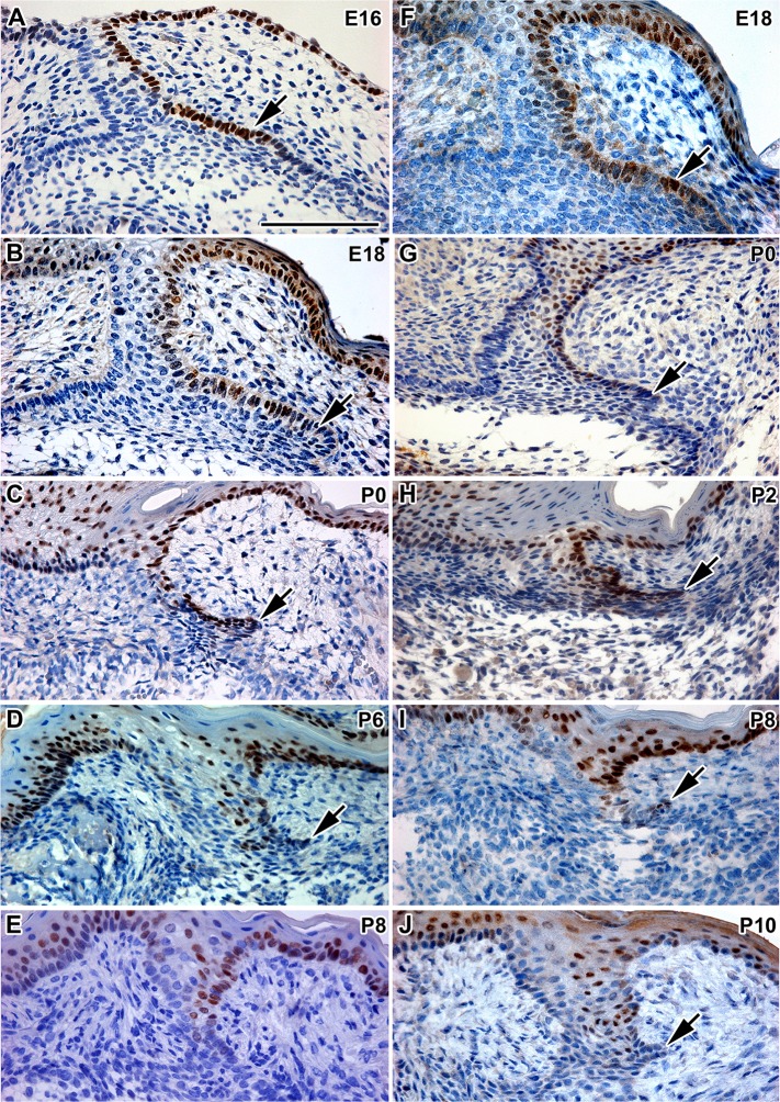
Fig 5. Sox2 protein expression in the dental stalk and rudimental successional dental lamina.
A: The epithelial thickening (arrow) on the lingual side of the tooth bell of M1 is Sox2-positive at E16. B: During late embryonic stages, a rudimental successional lamina as well as lingual side of the dental stalk is Sox2- positive. C, D: At postnatal stages, Sox2 is downregulated in the rudimental successional dental lamina of M1. E: At P8, the amount of Sox2-positive cells is decreased in the rudimental successional dental lamina. F: The epithelial thickening on the lingual side of the tooth bell of M2 is Sox2-positive at E18. G,H: Rudimental successional lamina is Sox2- positive at early postnatal stages (P0 and P2). I: Expression of Sox2 is downregulated in the rudimental successional dental lamina of M2 at P8. J: At P10, the amount of Sox2-positive cells is decreased in the dental stalk of M2. Sox2-positive cells are labeled by DAB (brown nuclei). Negative cells are counterstained by Hematoxylin (blue nuclei). Scale bar—100 μm

