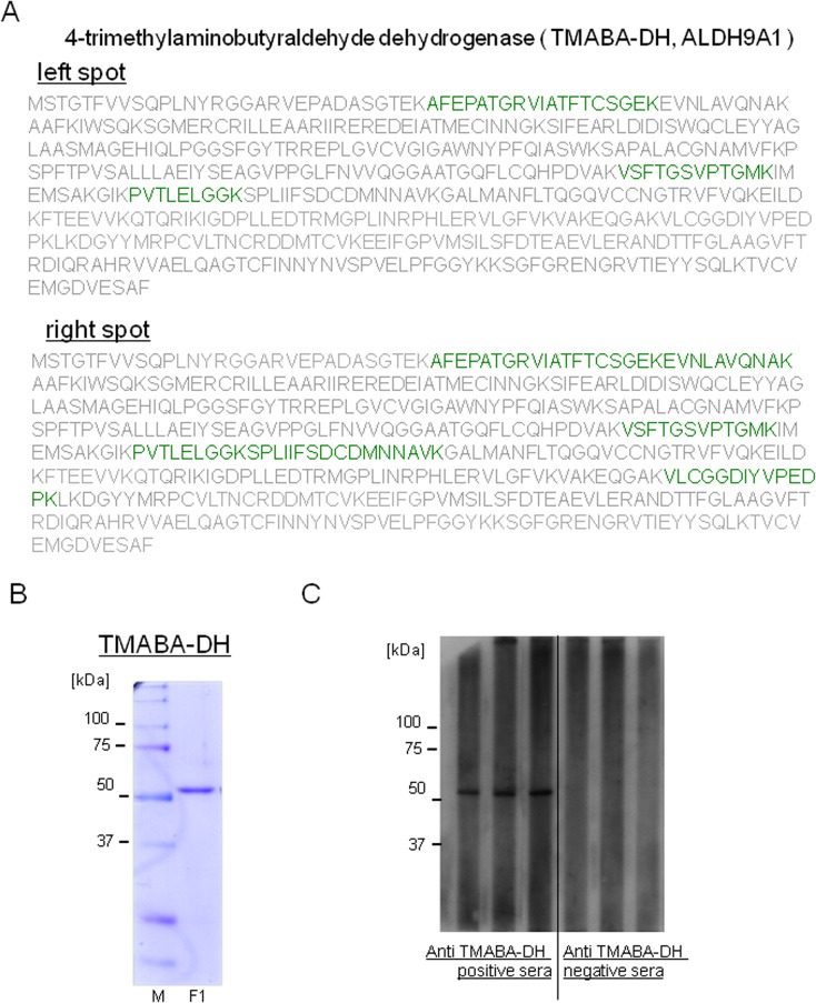
Fig 2. Identification of the autoantigen as TMABA-DH.
(A) Proteins in the spots indicated by arrows in Fig 1 were excised and subjected to lysylendopeptidase digestion. The resultant peptides were analyzed by LC-MS/MS as described under Materials and Methods. Proteins in both spots were identified as TMABA-DH (peptides identified using LC-MS/MS with >99% confidence are shown by green characters). (B) FLAG-tagged TMABA-DH was expressed in HEK293T cells, immunopurified using anti-FLAG M2 beads and competitively eluted using a solution containing FLAG peptide. An aliquot of the purified sample was analyzed using 9% SDS-PAGE followed by staining with coomassie brilliant blue. Molecular weight markers and their size in kilodaltons are shown on the left side of the figure. (C) Results of slot blot analyses of anti-TMABA-DH positive and negative sera are shown. Slot blot analyses were performed 5 times and the representative results are shown.

