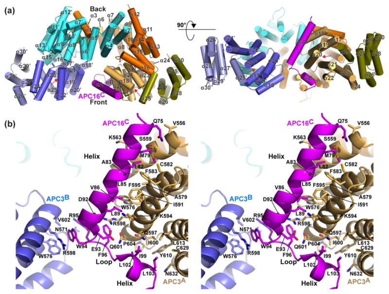
Fig. 4. Crystal structure of APC3-APC16: asymmetric interactions in a symmetric binding pocket.
a) Overall structure of APC3Δloop homodimer complex with APC16C. The APC3 domains are colored as in Fig. 2a. APC16 is colored magenta. Red arrow indicates the IR tail binding cleft.
b) Stereo view of close-up of the APC3-APC16 interface, showing side-chains mediating contacts.

