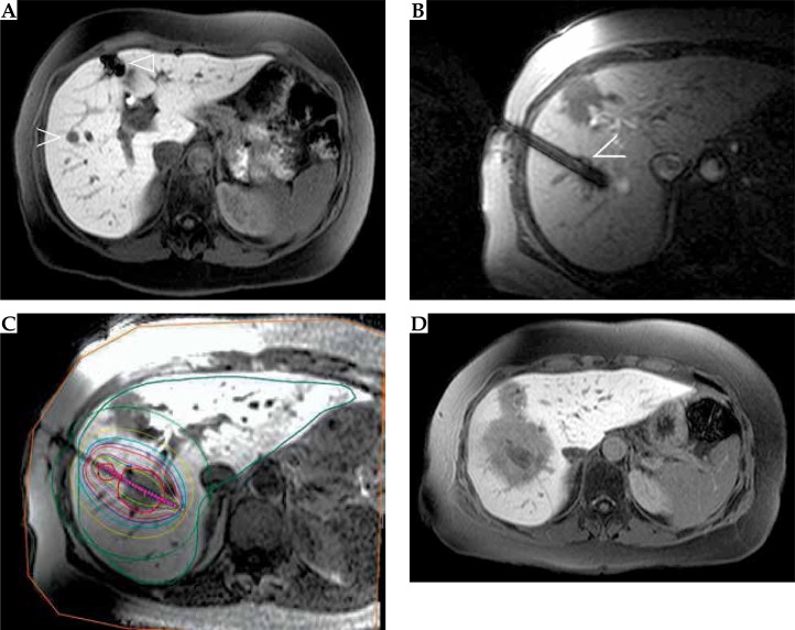
Fig. 1.
Local tumor control in a 58-year-old female patient with histological proven malignant melanoma metastases. A) Preoperative axial contrast-enhanced liver magnetic resonance imaging (MRI) (liver specific contrast agent Gd-EOB-DTPA) shows two metastases in segment V in the right liver lobe (open arrow), former partial resection of the liver segment IV (outlined arrow). B) Both metastases were treated by open MRI high-dose-rate brachytherapy using one catheter. C) Treatment planning and dosimetric analysis, tumor encircling isodoses (red line indicates 20 Gy). D) Follow-up contrast-enhanced liver MRI at 3 months shows the shrinking ablation zone with local control of the treated lesions

