Figure 3.

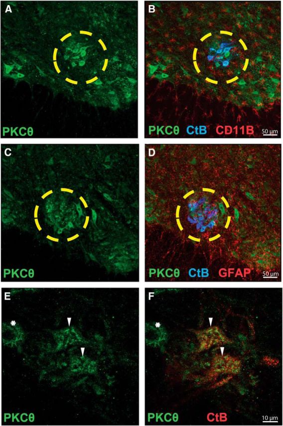
PKCθ is highly expressed in identified (CtB-positive) phrenic motor neurons. A, C, Representative confocal fluorescence images of ventral C4-C5 spinal sections from rats fluorescently labeled for PKCθ (Cell Signaling Technology, #9377, green). B, Colocalization of phrenic motor neurons (CtB, blue) and PKCθ fluorescence (green) is evident; in contrast, no colocalization could be appreciated between microglia (CD11b, red) and PKCθ (green) fluorescence. D, Colabeling for phrenic motor neurons (CtB, blue) and astrocytes (GFAP, red) reveals no expression of PKCθ in astrocytes. The yellow circle outlines the phrenic motor nucleus. E, Higher-magnification images of phrenic motor nucleus demonstrate PKCθ immunofluorescence (green). F, PKCθ immunofluorescence is present in phrenic motor neurons (CtB-positive, red; ▾), but also in unidentified putative neurons near the phrenic motor nucleus (CtB-negative, *).
