Figure 9.
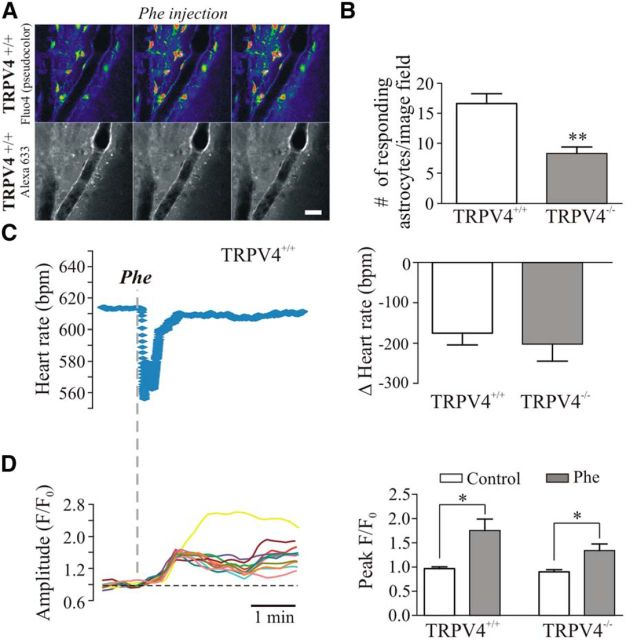
TRPV4 channels contribute to flow/pressure-induced astrocyte responses in vivo. A, 2PLMS images from a TRPV4+/+ mouse showing astrocytic Ca2+ activity (top) and the lining of the arteriole (bottom) following a systemic Phe injection. B, Summary data showing number of responding astrocytes following systemic Phe injection in TRPV4+/+ and TRPV4−/− mice. C, Representative trace showing changes in HR in response to Phe injection in a TRPV4+/+ mouse (left); summary data of Δ HR in TRPV4+/+ and TRPV4−/− mice (right). D, Representative trace showing changes in astrocytic Ca2+ activity in response to Phe injection in a TRPV4+/+ mouse (left); summary data showing changes in Ca2+ oscillation peak F/F0 amplitude in TRPV4+/+ and TRPV4−/− mice (right). *p < 0.05, **p < 0.01.

