
Fig. 7.
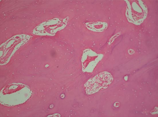
Photomicrograph of osteoma (Hematoxyline-eosin stain section in 100x magnification representing mature bone trabeculae with osteoblastic rimming and numerous osteocytes within lacunae in the represented area.)
Official websites use .gov
A
.gov website belongs to an official
government organization in the United States.
Secure .gov websites use HTTPS
A lock (
) or https:// means you've safely
connected to the .gov website. Share sensitive
information only on official, secure websites.
Photomicrograph of osteoma (Hematoxyline-eosin stain section in 100x magnification representing mature bone trabeculae with osteoblastic rimming and numerous osteocytes within lacunae in the represented area.)