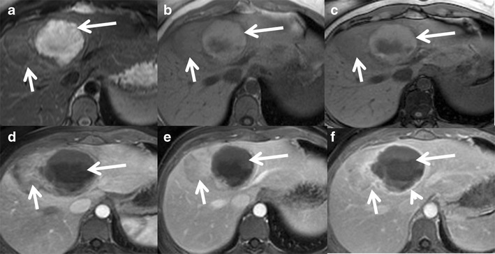
Fig. 12.

Hepatocellular adenoma with haemorrhage in a 40-year-old female on oral contraceptive pills. (a) Large heterogeneous lesion involving segment 8 and 4 of the liver showing a well-defined T2 hyperintense area along its medial aspect (long arrow) and T2 isointense area along its lateral aspect (short arrow). Axial T1 in-phase (b) and out-of-phase (c) MRI shows heterogeneous T1 hyperintensity in the medial portion (long arrow) without signal drop representing subacute haemorrhage. Axial post-contrast MRI shows heterogeneous mild-to-moderate enhancement of the lateral part of the lesion (short arrow) in the arterial phase (d) becoming isointense on venous (e) and delayed phases (f). The haemorrhagic component in the medial part of the lesion (long arrow) shows only peripheral capsular enhancement (arrowhead)
