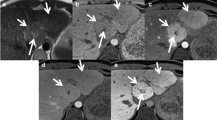
Fig. 2.

Classic FNH in a 35-year-old female with hepatobiliary-specific contrast (gadoxetate). (a) Axial T2W MRI shows two large isointense lesions in the left lobe of the liver (short arrows) with a T2 hyperintense central scar (long arrow). (b) Axial T1W MRI shows two large isointense lesions in the left lobe of the liver (short arrows) with a T1 hypointense central scar (long arrow). Axial post-contrast MRI with gadoxetate shows intense enhancement of the lesions (short arrows) in the arterial phase (c) becoming isointense on the portal venous phase (d) and persistent enhancement in the 20-min hepatobiliary phase (e). The central scar shows no enhancement (long arrow)
