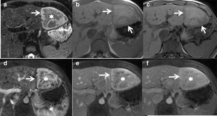
Fig. 6.

A 30-year-old female with incidental detection of a hypoechoic nodule on ultrasound. (a) Axial fat-saturated T2W MRI shows an iso- to hyperintense lesion (asterisk) in the left lobe of the liver with a peripheral T2 hyperintense rim (arrow) representing the atoll sign. Axial T1 in-phase (b) and out-of-phase (c) MRI shows no drop in signal (arrows). (d) Axial post-contrast MRI in the arterial phase shows moderate heterogeneous central enhancement (asterisk) and peripheral rim enhancement (arrow). Axial post-contrast MRI in the venous phase (e) and delayed phases (f) shows persistent enhancement (asterisk) with delayed enhancement of the peripheral rim (arrow). Surgical resection confirmed the diagnosis of inflammatory hepatocellular adenoma
