Fig. 4.

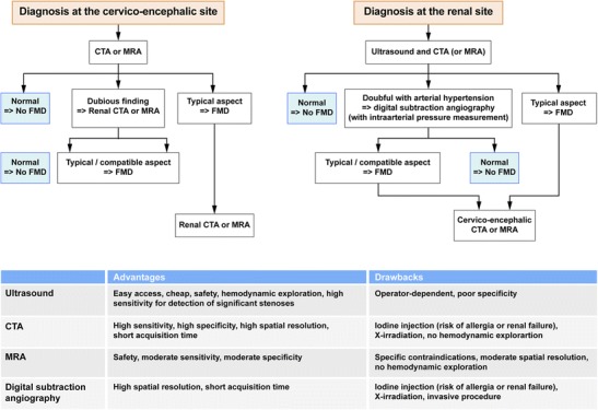
Flow chart illustrating imaging algorithms in case of suspected DFM for both the renal or cervico-encephalic level. The table shows the advantages and drawbacks of the different diagnostic tests

Flow chart illustrating imaging algorithms in case of suspected DFM for both the renal or cervico-encephalic level. The table shows the advantages and drawbacks of the different diagnostic tests