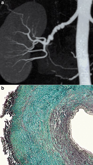
Fig. 2.

Example of medial FMD lesions. a Renal CTA with coronal plane MIP reconstruction, showing the typical “string-of-beads” aspect of the right renal artery, in favour of medial FMD. b Colour histological slide of medial FMD showing extensive medial fibrosis. Intima and adventitia are normal
