
Figure 3. Ion adsorption at mica-water interface.

(a) Surface Charge calculated from ζ potential measurements (circles) vs. concentration of solutions of NaCl (red) and CaCl2 (blue) at pH 6. Solid lines: surface complexation model predictions. Blue triangles: AFM data from25; blue squares24, red triangles23, red squares32: surface forces apparatus measurements. The charge density is normalized by the characteristic scale  arising from the Poisson-Boltzmann equation,
 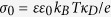 , where
  is the Debye parameter. (b) AFM images of mica-water interface showing the characteristic hexagonal lattice of mica in 100 mM NaCl solution (left), and a rectangular symmetry caused by (presumably hydrated) adsorbed Ca2+ ions in 100 mM CaCl2 (right). Insets: filtered zoomed views with overlaid lattice structure (top) and Fast Fourier Transform image of the same data (bottom). c Gray scale encoded contact angle vs. pH and CaCl2 concentration. Top: model prediction; bottom: experimental data. Symbols (x: θ < 2°) and numbers: experimental data same as Fig. 1b with interpolated gray scale. Smoothed lines are guides to the eye based on the experimental datapoints.