Figure 2.
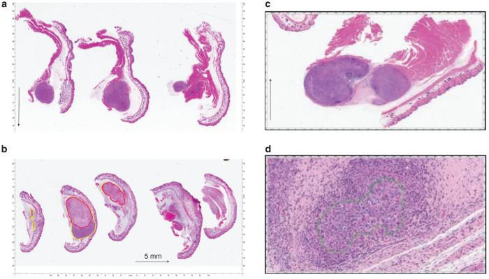
H & E staining of hind limb tumors. (a) Untreated control: micrograph showing viable Panc-1 human tumor xenograft from untreated control (black arrow denotes 5 mm in size). (b) 7 day posttreatment survival. Micrograph showing Panc-1 human tumor xenograft 7 days after IRE treatment. Using Aperio ScanScope xenograft is outlined with yellow. Nonviable tumor is outlined with red (black arrow denotes 5 mm in size). (c) 21 day posttreatment survival: no viable tumor present, tumor cells are surrounded by fibrosis, and mononuclear cells. There is viable muscle adjacent to tumor nodule (black arrow denotes 5 mm in size). (d) 21 day posttreatment survival. Shows tumor cells surrounded by chronic inflammation.

