Figure 3.
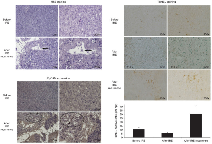
Histological examination of incomplete recurrences. (i) Hematoxylin and eosin staining: areas of infiltration and necrosis (arrows) amid viable tumor cells in the recurrences (lower plate) on 100 and 200× original magnification. (ii) Immunohistochemical staining of recurrences with EpCAM antibody: higher EpCAM expression in recurrent tumors (lower plate) compared to original Panc-1 tumors (white arrows lower panels and the example of increased EpCAM expression as defined by the deep dark brown/black cells). (iii) Higher apoptotic turnover (TUNEL staining: dark brown arrows) in IRE recurrences (middle plate) compared to original Panc-1 tumors (top plate) and completely ablated tumors (bottom plate). There is a higher EpCAM expressed cancer cells in the recurred tumors compared to the tumor of original Panc-1 cells inoculation by immunohistochemical staining (EpCAM see circles). All these features indicate that the recurrent tumor may be more aggressive than the tumor of original Panc-1 cells inoculation.

