Figure 4.
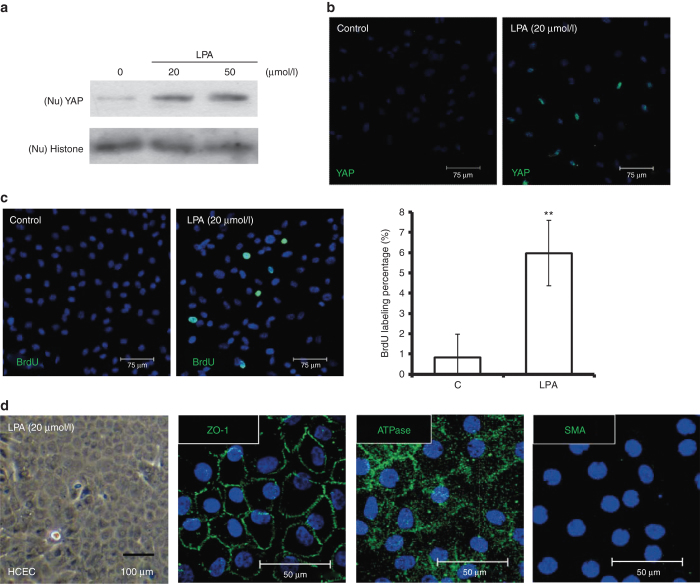
LPA enhances nuclear levels of YAP and promotes proliferation in contact-inhibited HCECs. (a) The stimulatory effects of LPA on nuclear levels of YAP in contacted-inhibited HCECs were evaluated by western blotting assays. HCEC monolayers were serum starved for 24 hours and incubated in LPA at different doses for an additional 4 hours. The nuclear proteins were extracted from each experimental group and fractionated on 10% SDS–PAGE gels (5 μg/lane). After transfer, the membranes were blotted with an antibody against either YAP or histone as a loading control. (b) The distribution of nuclear YAP (green) was detected in immunofluorescein assays in contacted-inhibited HCECs 4 hours after the addition of 20 μmol/l LPA. (c) Cell proliferation was examined via BrdU labeling, and the BrdU labeling (green) in HCEC monolayers treated with 20 μmol/l LPA was significantly higher than in HCEC monolayers treated with phosphate-buffered saline (n = 3; **P < 0.01). (d) HCECs treated with LPA (20 μmol/l) showed a hexagonal morphology under phase microscopy, with a normal immunostaining pattern for ATPase and ZO-1, but without any expression of SMA, indicating no evidence of endothelial–mesenchymal transition. The cell nuclei were counterstained with Hoechst 33342 (blue). LPA, lysophosphatidic acid; SMA, smooth muscle actin; YAP, yes-associated protein.

