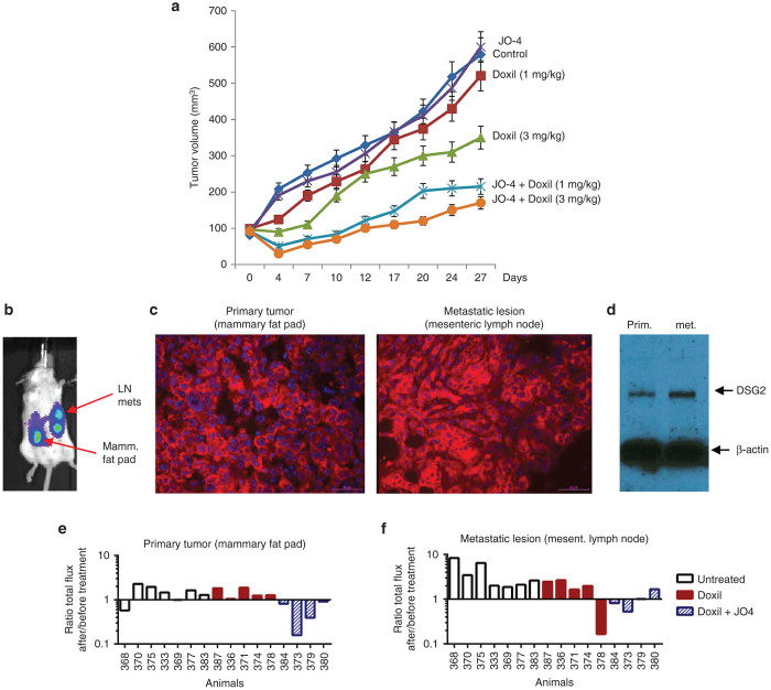
Figure 2.
Efficacy studies with JO-4 and Doxil in mouse models. (a) Efficacy study in mammary fat tumor pad model using primary ovarian cancer (ovc316) cells. Treatment was started when tumors reached a volume of 100 mm3. Mice were injected intravenously with Doxil (1 or 3 mg/kg) alone or in combination with 2 mg/kg JO-4. Treatment was repeated weekly. N = 10. (b–f) Studies in the spontaneous metastasis model based on MDA-MB-231-luc-D3H2LN cells. (b) In vivo luciferase imaging of a representative animal. The primary tumor in the mammary fat pad forms metastasis in regional lymph nodes (LN), e.g., pancreatic and mesenteric lymph nodes. (c) DSG2 immunofluorescence analysis of sections from the primary tumor and mesenteric lymph node metastases. DSG2 signals are in red and appear to be membrane associated. Nuclei are stained in blue. (d) Western blot analysis of primary tumor and metastases with DSG2 antibodies. β-actin is used as a loading control. A representative animal is shown. (e,f) Comparison of in vivo luciferase signal in the primary tumor (e) and metastasis (f) 1 day before and 5 days after treatment with Doxil or JO-4 + Doxil. Shown is the total flux of the primary tumor or metastasis ROI after treatment divided by the ROI before treatment. Shown are averages of imaging sequences of five images each. The difference between the Doxil and Doxil+JO4 groups is significant (P < 0.01) for both the primary tumor and the metastases. The difference between the untreated and Doxil groups is not significant.

