Figure 1.

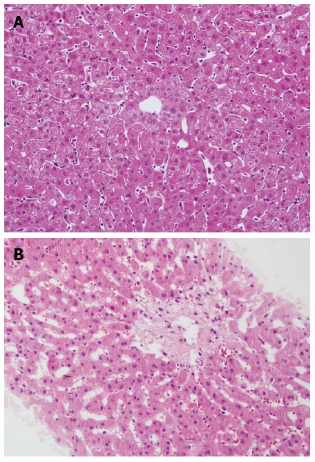
Biopsy of the donated graft at the time of transplantation was normal (HE, × 200) (A) and liver biopsy showed sinusoidal congestion and fibrosis of centrilobular veins at 88 d after transplantation (HE, × 200) (B).

Biopsy of the donated graft at the time of transplantation was normal (HE, × 200) (A) and liver biopsy showed sinusoidal congestion and fibrosis of centrilobular veins at 88 d after transplantation (HE, × 200) (B).