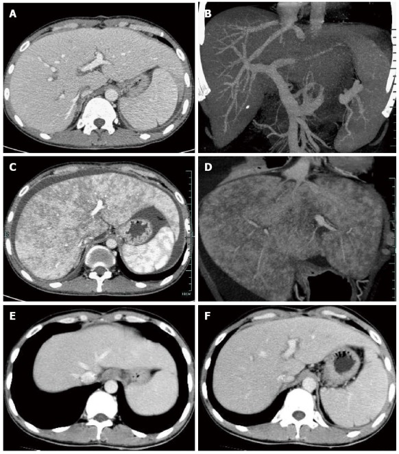
Figure 2.

Computed tomography revealed excellent reconstructed blood flow in the hepatic veins, portal vein and hepatic artery at 14 d after transplantation (A, B), enlarged liver with patchy enhancement, obscure hepatic veins and massive ascites at 80 d (C, D), and normal radiologic presentations with resolved ascites and patchy enhancement, and recovered hepatic vein flow at 120 d (E, F).
