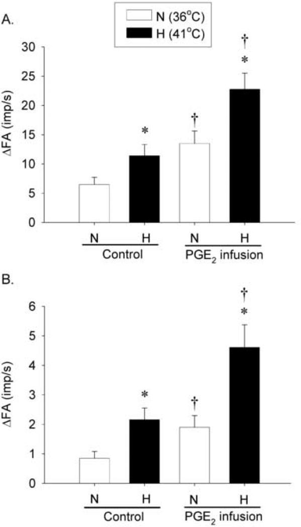
Fig. 4. Effects of PGE2 on the baseline activity and responses of pulmonary C fiber to right-atrial bolus injections of capsaicin and adenosine.
A, average responses to capsaicin injection (0.5-1.0 μg/kg, n = 18); B, average responses to adenosine injection (170 μg/kg, n = 12). Average peak responses of pulmonary C fibers to right-atrial injections of capsaicin and adenosine were measured with or without PGE2 infusion (3 μg/kg/min, 3 min) at 2 different levels of Tit (N: 36°C, open bars; H: 41°C, closed bars). Each of the chemicals (0.15 ml) was slowly injected into the right atrium as a bolus. ΔFA represents the difference between the peak FA (average over 2-sec interval for capsaicin and 10-sec interval for adenosine injection) and the baseline FA (average over 60-sec interval) in each fiber. *, significantly different (P < 0.05) from the corresponding data at normal Tit (36°C); †, significantly different (P < 0.05) from the corresponding data at control (before PGE2 infusion). Data are mean ± S.E.M.

