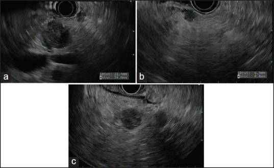
Figure 1.

Endoscopic ultrasound images revealing multiple well-demarcated, hypoechoic pancreatic lesions ((a) 24.6 mm × 21.4 mm mass in the body of the pancreas, (b) 6.9 mm × 6.6 mm nodule in the body, and (c) 14.1 mm × 11.7 mm and 10.6 mm × 7.2 mm masses in the pancreatic head)
