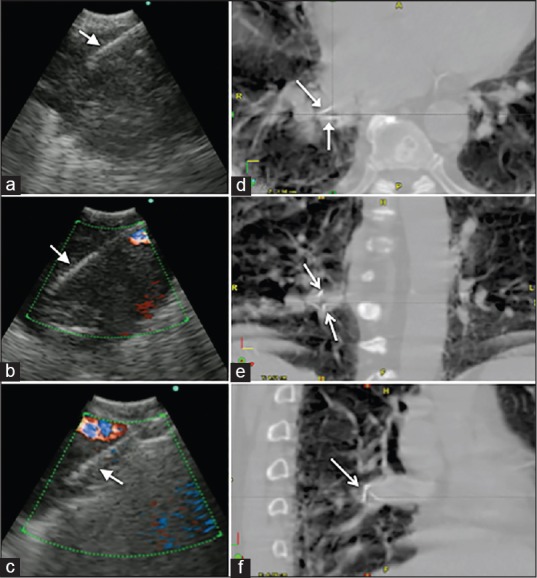
Figure 2.

(a-c) Convex probe endobronchial ultrasound images showing the EBUS needle (arrows) at different locations inside the tumor representing where the three fiducials markers were deployed. (d-f) Images from the stereotactic body radiation therapy planning scan showing the fiducial markers inside the tumor (arrows)
