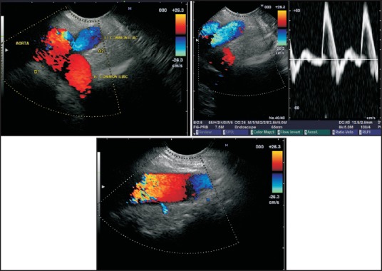
Figure 34.

Imaging of the lowest part of the descending aorta is possible from the rectum. The scope should be positioned at about 20 cm distance (~rectosigmoid). From the rectosigmoid junction the vessel coming towards the probe is the left common iliac artery. The small lumbar arteries can be seen joining the aorta by clockwise and anticlockwise rotation
