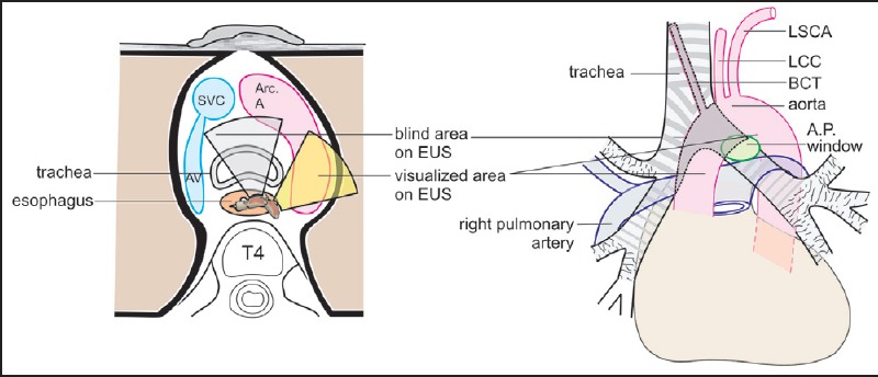
Figure 8.

The shaded area shows the blind areas of imaging during endoscopic ultrasound. The upper part of ascending aorta, the brachiocephalic trunk and part of the arch of aorta lie in front of trachea and cannot be visualized by endoscopic ultrasonography. SVC = Superior vena cava, AV = Azygos vein, Arc. A = Arch of aorta, T4 = Fourth thoracic vertebra, LSCA = Left subclavian artery, LCC = Left common carotid artery, BCT = Brachiocephalic trunk, AP window = Aortopulmonary window
