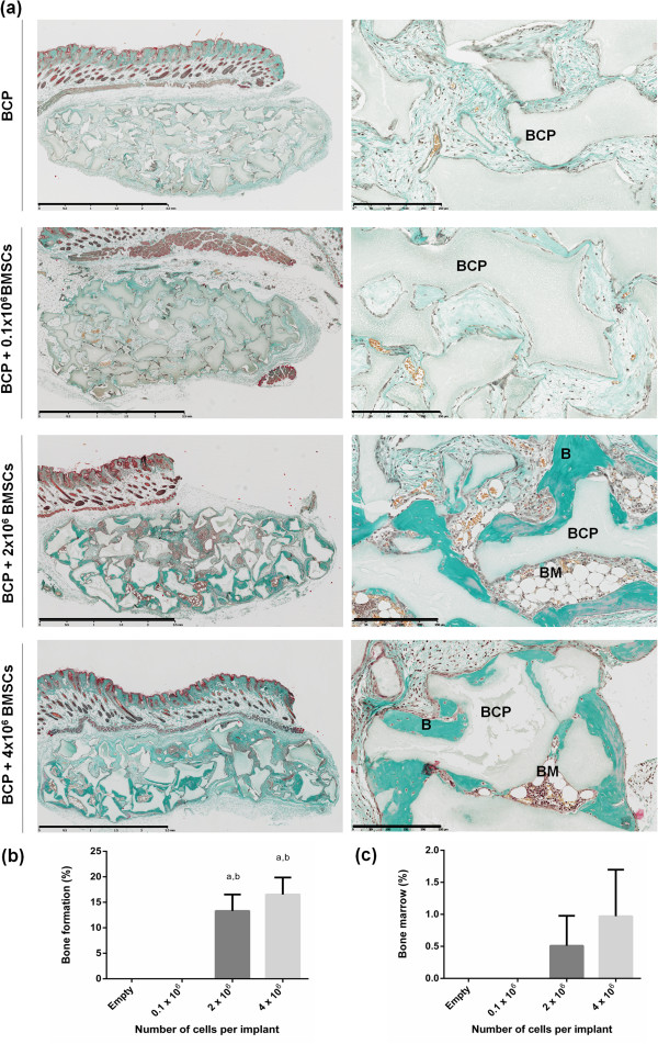
Figure 2.

Optimal cell dosage for ectopic bone formation. (a) Masson trichrome staining shows biphasic calcium phosphate biomaterial (BCP, gray) in contact with newly formed bone (B, green). Mature bone marrow territories (BM) were present after 8 weeks. Scale bars: 2.5 mm and 250 μm for images in the left and right columns respectively. (b) Histomorphometry revealed significantly more bone in the 2 × 10⁶ to 4 × 10⁶ cell groups compared with the empty scaffolds (a P <0.02) and compared with the 0.1 × 10⁶ cell group (b P <0.02). (c) Bone marrow territories were present only in the 2 × 10⁶ and 4 × 10⁶ cell groups, but there was no statistical difference between groups. BMSCs, bone marrow stromal cells.
