
Figure 4.
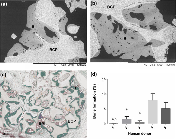
Ectopic bone formation of freshly transported batches of bone marrow stromal cells. (a), (b) In backscattered electron imaging, the biphasic calcium phosphate (BCP) ceramic and mineralized bone can be differentiated by their relative gray densities. The BCP particles were surrounded by a well-mineralized lamellar bone tissue in the BCP + BMSCs group after 8 weeks. (c) Masson trichrome staining showed bone formation (B) at the periphery of the explants that contained bone marrow compartments (BM). (d) Histomorphometry of Masson trichrome staining sections revealed significant differences in the bone induction capacity between human donors. aStatistically lower compared to Donor 4 (P <0.05). bStatistically lower compared with to Donor 5 (P <0.05). BMSCs, bone marrow stromal cells.