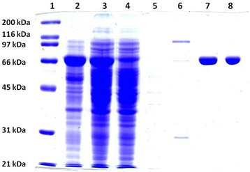
Fig. 1.

Analysis of the expression and purification of the recombinant α-l-fucosidase from P. thiaminolyticus by SDS-PAGE. Electrophoresis was carried out in 10 % polyacrylamide gel and proteins were visualized by Coomassie Brilliant Blue R-250. Line 1- SDS-PAGE Molecular Weight Standards, Broad Range, line 2 - cells of E.coli BL21 (DE3) after expression of α-l-fucosidase iso2, line 3 - supernatant after disintegration of cells after expression, line 4 - proteins which did not bind to the Ni-NTA agarose after the application of the supernatant sample, line 5 - fraction after wash in the binding buffer with 10 mM imidazole, line 6 - fraction after wash in the binding buffer with 40 mM imidazole, lines 7 and 8 - recombinant α-l-fucosidase iso2 from P. thiaminolyticus eluted by the binding buffer with 250 mM imidazole and desalted by gel filtration on the column PD10
